Research in the Vanderbilt Mass Spectrometry Research Center (MSRC) spans the full spectrum of mass spectrometry development and applications. From proteomics to metabolomics to structural characterization and advanced multimodal imaging mass spectrometry, the MSRC is an internationally recognized hub for both technology innovation and biomedical discovery.
Interested in learning more about research opportunities in MSRC labs? Reach out to researchers directly or email us at MSRC@vanderbilt.edu
Publications
A lipid atlas of the human kidney
Glycolipids implicated as mediators of clinically visible retinal pigment epithelial migration in age-related macular degeneration
Spatial patterns of hepatocyte glucose flux revealed by stable isotope tracing and multi-scale microscopy
High-Specificity and Sensitivity Imaging of Neutral Lipids Using Salt-Enhanced MALDI TIMS
Preserving full spectrum information in imaging mass spectrometry data reduction
Species-specific components of the Helicobacter pylori Cag type IV secretion system
In situ molecular profiles of glomerular cells by integrated imaging mass spectrometry and multiplexed immunofluorescence microscopy
Endogenous Aquaporin-0 Lipid Binding in Ocular Lens Tissue via Native Mass Spectrometry
A thiouracil desulfurase protects Clostridioides difficile RNA from 4-thiouracil incorporation, providing a competitive advantage in the gut
Cutting Edge Technology
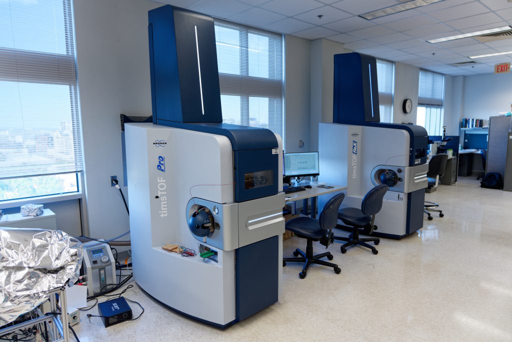
The MSRC is a leader in technology innovation, developing new mass spectrometry instrumentation and methods and hosting one of the most advanced collections of instrumentation in the world. The center houses 45 state-of-the-art mass spectrometers spanning all major vendors and platforms, alongside orthogonal technologies including highly multiplexed immunofluorescence microscopy and spatial transcriptomics systems. Together, these resources provide unparalleled capabilities for multi-omics and spatial biology.
Schey Research Laboratory
The focus of the Schey lab is on the development and application of mass spectrometry to understand fundamental biochemical processes in health and disease. Areas of technology development include: imaging mass spectrometry, membrane protein analysis, and proteomics. Areas of application include: protein aging, ocular lens biochemistry and retina diseases such as age-related macular degeneration. State-of-the-art instrumentation and methods, such as hydrogen-deuterium exchange MS, imaging mass spectrometry, native mass spectrometry, top-down mass spectrometry, and single cell proteomics, are employed to examine protein-protein interactions, post-translational modifications, protein crosslinking, spatially-resolved proteomics/lipidomics, and cell aging.
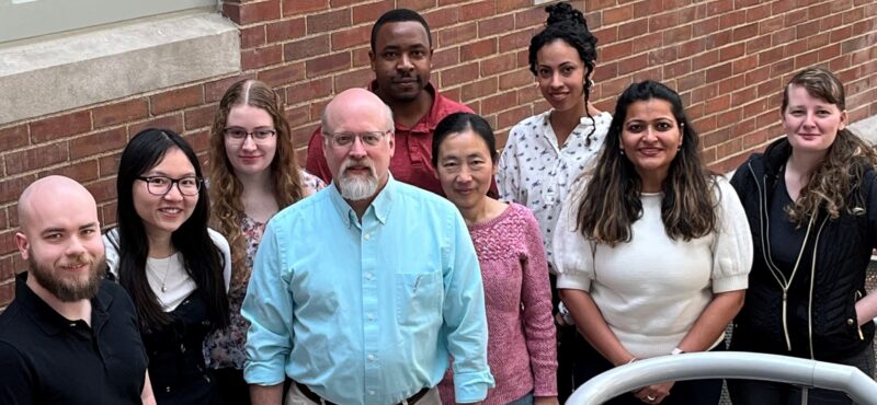
Spraggins Research Laboratory
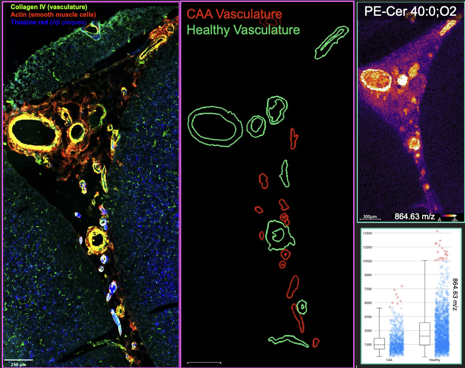 The Spraggins Lab pioneers next‐generation imaging mass spectrometry and integrated multimodal molecular imaging to map molecular landscapes in tissues—spanning metabolites, lipids, proteins, and genes—with spatial resolution from single cells to whole organs. Their work blends high-performance IMS instrumentation, computational tools, and complementary modalities (e.g. multiplexed immunofluorescence, spatial transcriptomics) to build molecular atlases and to illuminate disease biology in Alzheimer’s, kidney disease, infection, and more.